Three-dimensional micro X-ray fluorescence analysis (3D-µRFA)
Application of a 3D-µRFA benchtop spectrometer for non-destructive, depth-resolved elemental analysis
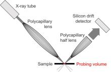
Three-dimensional micro X-ray fluorescence analysis (3D-µRFA) has only been used as a benchtop spectrometer for around two decades and has since been the subject of research for a wide range of applications. The depth sensitivity of the spectrometer is based on a defined three-dimensional examination volume, which results from a confocal arrangement of two polycapillary lenses in the excitation and detection channel. The spectrometer used at the institute is a modified µRFA table-top spectrometer (M4 TORNADO) from Bruker Nano GmbH. The setup is based on the research results of the Kanngießer group (TU Berlin, Institute of Optics and Atomic Physics). As a result of the modification, a second polycapillary lens was installed perpendicular to the first in front of a 60 mm2 silicon drift detector.
To validate the 3D-µRFA setup, various parameters such as penetration depth, matrix effects, absorption effects and dimensions of the examination volume are evaluated. Among other things, self-synthesised layered polymer standards with different concentrations of selected elements are used for this purpose.
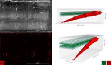
There are various areas of application for 3D micro-XRF, but ideally these are real samples with a light matrix, as this is where the greatest penetration/information depth can be achieved. One example of this is the investigation of the antibacterial effect of sponges and spongin scaffolds, in which the three-dimensional distribution of copper for a sponge that was previously treated with copper-containing wastewater was mapped using 3D-µRFA. The formation of a sponge-atacamite composite was detected. By dissolving out the copper-containing biominerals, it is possible to regenerate the sponge and thus reuse it for the purification of industrial wastewater in several cycles. (Cooperation partner: Prof. Dr rer. nat. habil. Hermann Ehrlich)
Another important field of application is the three-dimensional analysis of minerals, particularly with regard to the characterisation of mineral inclusions. The non-destructive nature of 3D micro-XRF allows the chemical phase composition to be analysed and the corresponding phase identification to be carried out while maintaining the integrity of the mineral inclusion isolation. A wide variety of mineral systems with different phase and inclusion compositions, such as quartz, fluorite, zircon, garnet and wolframite, were analysed for this purpose. (Cooperation partner: Dr Axel Renno) Other promising areas of application for 3D micro-XRF include archaeometry of historical objects and the three-dimensional analysis of plant tissue.
Publications
Tsurkan, D.; Simon, P.; Schimpf, C.; Motylenko, M.; Rafaja, D.; Roth, F.; Inosov, D. S.; Makarova, A. A.; Stepniak, I.; Petrenko, I.; Springer, A.; Langer, E.; Kulbakov, A. A.; Avdeev, M.; Stefankiewicz, A. R.; Heimler, K.; Kononchuk, O.; Hippmann, S.; Kaiser, D.; Viehweger, C.; Rogoll, A.; Voronkina, A.; Kovalchuk, V.; Bazhenov, V. V.; Galli, R.; Rahimi-Nasrabadi, M.; Molodtsov, S. L.; Joseph, Y.; Vogt, C.; Vyalikh, D. V.; Bertau, M.; Ehrlich, H. (2021) Extreme Biomimetics: Designing of the First Nanostructured 3D Spongin-Atacamite Composite and ist Application. Advanced Materials, 2101682 (Early View)
Heimler, K.; Gottschalk, C., Vogt, C.; Confocal Micro X-Ray Fluorescence Analysis for the Non-Destructive Investigation of Structured and Inhomogeneous Samples (Critical Review), ABC 2023, 415, 5083-5100 https://doi.org/10.1007/s00216-023-04829-x
Cooperation partners
Prof. Dr. rer. nat. habil. Hermann Ehrlich, Chair of Biomineralogy and Extreme Biomimetics
Dr Axel Renno, Department of Analytics at the Helmholtz Centre Dresden-Rossendorf
Prof B. Kanngießer, TU Berlin


