Sweet flavours and healthy ingredients make strawberries one of the world's most popular berries. The complex biochemical compounds that determine the flavour and nutritional value of a strawberry - known as biomarkers - can only be determined in detail by experts using modern analyses. A team from TU Bergakademie Freiberg, together with Serbian researchers, has now analysed different strawberry varieties for the first time using ultra-high resolution mass spectrometry and examined how the biomarkers are distributed in the fruit. The chemists discovered that most of the biomarkers are found in the red skin of the strawberries. The team recently published the results in the journals "Wiley Analytical Science" and "Plants".
In the strawberry skin, for example, the team of researchers led by Dr Jan Zuber from TU Bergakademie Freiberg identified the biomarker pelargonidin-3-O-malonyglucoside. "This molecule has an antioxidant effect. In the human body, these chemical compounds can bind free radicals and thus have an anti-inflammatory and vasoprotective effect," explains the research associate at the Institute of Analytical Chemistry. The strawberry varieties analysed also contain other biomarker molecules such as organic acids, for example citric acid, or sugars such as glucose. Depending on the concentration and ratio of these biomarkers, the flavour of the respective strawberry variety and its nutritional quality are determined. The majority of these biomarkers are also found in the strawberry skin.
Thin strawberry slices for detailed analyses
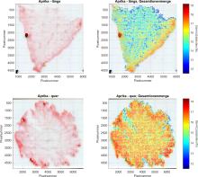
The fact that the team was able to detect the concentration of biomarkers in the fruit so precisely is due to the high-tech analysis device used by the researchers to examine the thin strawberry slices, the ultra-high-resolution mass spectrometer. Here, the biomarkers are first converted into charged particles, known as ions, by a laser and then separated according to their mass and charge number. "Our analyses made it possible for the first time to distinguish between different biomarkers whose masses differ from each other only to the sixth decimal place." The biomarkers are represented with a colour-coded image of the measured signals of the wafer-thin strawberry slice. "This clearly shows that the biomarkers relevant to the taste and quality of the fruit are found in particularly high concentrations in the skin of the strawberry," says Zuber.
Targeted breeding of strawberries
Together with researchers from the Serbian University of Belgrade, Dr Jan Zuber's team used this method to investigate a total of 25 new strawberry varieties. "As these biomarkers are decisive for the flavour and also the quality of strawberries, it is interesting for the breeding of strawberry varieties to know exactly where the biomarkers are located and in what concentration," says the chemist. "These findings can be used to select fruit varieties for production that are as high yielding and rich in nutrients as possible. On the other hand, the results also help to assess how new fruit varieties will be accepted by end consumers."
Background: Analysing samples with the mass spectrometer
The sample used for the analysis is only a few micrometres thick. It is prepared for measurement in the mass spectrometer using a special cutting device (cryo-microtome). The wafer-thin disc is then coated with a UV-active matrix, which is necessary for the conversion of the various biomarkers into the corresponding ions. The researchers then place a measurement grid over this preparation. Each point of this grid is now excited with the laser of the mass spectrometer and the resulting ions are detected. Thanks to the ultra-high resolution mass spectrometer, even the smallest differences in mass between the biomarker ions can be detected and used for differentiation. The information obtained is then evaluated and visualised using distribution images to show which biomarkers can be detected in which areas of the thin sections.
The ultra-high-resolution measurements were carried out by the German-Serbian team as part of a project funded by the German Academic Exchange Service DAAD (reference number: 57602001).
